- Your cart is empty
- Continue Shopping
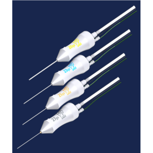
Dry Amniotic Membrane

Product Description
Preserved human amnion has been successfully used as a biological bandage, promoter of epithelialisation, inhibitor of inflammation and angiogenesis, as well as a carrier for ex vivo cultured limbal stem cells. The purpose of this Histologically the amnion is a 0.02 mm to 0.5 mm five-layered membrane, composed of three basic layers.
Epithelial monolayer:
The epithelium consists of a single layer of cuboidal cells with a large number of microvilli on the apical surface. The basement membrane is a thin layer composed of a network of reticular fibers. Histochemically the basement membrane closely resembles that of the conjunctiva. The compact layer contributes to the tensile strength of the membrane. The fibroblast layer is the thickest layer of the AM made up of a loose fibroblast network. The outermost layer of the amnion is the spongy layer. The basement membrane is one of the thickest membranes found in human tissue. This layer is resistant to current cryopreservation techniques. The structural integrity, transparency and elasticity of the amniotic basement membrane make it currently the most widely accepted tissue replacement for ocular surface reconstruction. It is known to promote epithelial cell migration, adhesion and differentiation. It is an ideal substrate for supporting the growth of epithelial progenitor cells by prolonging their lifespan, maintaining their clonigenicity and preventing epithelial cell apoptosis. This action explains why AMT facilitates epithelialisation for PED with stromal ulceration. In tissue cultures AM supports epithelial cells grown from explant cultures and maintains their normal morphology and differentiation. The resultant cultured epithelium can be transplanted with the AM to reconstruct damaged corneas. The AM can be used to promote non-goblet cell differentiation of the conjunctival epithelium.
Thick basement membrane:
The basement membrane of the AM, cornea and conjunctiva contain collagen types IV, V and VII, in addition to fibronectin and laminin. Though the laminins are very effective in facilitating corneal epithelial cell adhesion Type V collagen helps in the epithelial cell anchorage to the stroma.
Avascular, hypocellular stromal matrix:
AM produces basic fibroblast, hepatocyte and transforming growth factor (TGF). These growth factors can stimulate epithelialisation and modulate proliferation and differentiation of stromal fibroblasts. The AM stromal matrix, rich in fetal hyaluronic acid suppresses TGF B signaling, proliferation and myofibroblastic differentiation of normal corneal and limbal fibroblasts as well as normal conjunctival and pterygium fibroblasts. This action explains why AMT helps reduce scars during conjunctival surface reconstruction, prevents recurrent scarring after pterygium removal and reduces corneal haze following photorefractive keratectomy. The stromal matrix also suppresses expression of certain inflammatory cytokines that originate from the ocular surface epithelia, including interleukin 1a, IL -2, IL-8, ?, tumor necrosis factor-, basic fibroblast growth factor and platelet derived growth factor. The AM attracts and sequesters inflammatory cells infiltrating the ocular surface and contains various forms of protease inhibitors. This may explain some of its anti-inflammatory properties.
Indications:
- Conjunctival surface reconstitution
- Pterygium surgery.
- Chemical & burn injury
- Cicatrizing conjunctivitis
- Ocular surface squamous neoplasia(ossn)
- Leaking blebs
- Filtering surgery
- Symblepharon release
- Fornix formation
- Socket reconstruction
- Conjunctivochalasis
| Weight | 0.5 kg |
|---|


Reviews
There are no reviews yet.